

Anna Laura Giacomin
Camposampiero Hospital, Italy
Title: Arrested foveal development and low visual function in babies with history of retinopathy of prematurity
Biography
Biography: Anna Laura Giacomin
Abstract
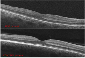
Introduction: Retinopathy of prematurity (ROP) can lead to blindness due to retinal detachment, but also premature infants with a normal apparent retina can develop subnormal visual acuity. Foveal depression and inner retinal layers develop mainly before the 40 week of gestational age (GA), while outer retinal layers develop mainly post birth. The purpose of our study was to correlate inner and outer foveal structural alterations with visual function and to correlate these alterations with GA, body weight (BW), and stage of ROP in preterm infants with ROP regressed spontaneously or after laser treatment.
Methods: Thirty eyes of 15 premature children aged 4 to 9 years with previous ROP underwent a complete ophthalmological and orthoptic evaluation, spectral domain OCT and angio-OCT (Canon OCT-HS100). The parameters evaluated with SDOCT in the fovea were: Total retinal thickness, inner retinal layers thickness, Henle’s layer thickness, outer nuclear layer thickness, external limiting membrane integrity, ellipsoid zone and outer segment tips zone integrity. Foveal avascular zone and foveal intra-retinal vascular plexus were evaluated with ANGIO-OCT. Other parameters evaluated were: Best corrected visual acuity (BCVA), refraction after cycloplegia. 10 eyes of 5 normal term children served as controls.
Results: Total central foveal thickness, inner retinal layer thickness, Henle’s layer thickness are significantly higher compared to term children. Outer nuclear layer thickness is lower compared to term children. Impaired visual function is correlated to impaired outer retinal layers structure. These changes are correlated to GA, BW, and stage of ROP.
Conclusions: A correct macular development at birth period is crucial for normal visual function. An impaired foveal cell migration and development can lead to a subnormal visual function in children with history of ROP. Further studies are necessary to understand the causes of foveal structure changes in preterms and the influence of macular edema of prematurity.